
X-Ray Interaction: Photoelectric Effect, Compton Scattering
Understand X-ray photoelectric effect and Compton scattering physics to design detectors and optimize contrast in medical imaging.
Contents & prerequisites
Why X-Ray Interaction Matters in Medical Devices
Role of photon-tissue interaction in diagnostic imaging quality
Every radiograph, fluoroscopic frame, and CT slice is a map of how X-ray photons were absorbed or deflected as they passed through tissue. The detector never "sees" anatomy directly — it sees the surviving photon fluence pattern after millions of individual photon-electron interactions have occurred along each ray path. If you are designing a detector, choosing scintillator material, or setting exposure protocols, you are implicitly working with two dominant physical processes: the photoelectric effect and Compton scattering. Everything downstream — contrast-to-noise ratio, patient dose, scatter artifacts, Hounsfield unit stability — traces back to the relative probability of these two interactions at a given photon energy and tissue composition.
How interaction mechanisms determine image contrast and signal
Photoelectric absorption removes a photon from the beam entirely and deposits its energy locally; Compton scattering redirects the photon with reduced energy and sends it off in a new direction, often toward the detector from an angle that carries no useful spatial information. Image contrast depends on the difference in attenuation between adjacent tissues, and that difference is driven almost entirely by how much photoelectric absorption each tissue type experiences, since Compton interaction probability is comparatively insensitive to tissue type. Understanding which mechanism dominates where is therefore the first design decision in any X-ray imaging system.
Energy ranges relevant to clinical radiography and fluoroscopy
Diagnostic radiography typically operates between 40 and 150 kVp (tube voltage), producing polyenergetic spectra with effective energies in the 20–80 keV range after filtration. Mammography sits at the low end (roughly 25–35 kVp, with most photons between about 15 and 30 keV), fluoroscopy typically runs 60–120 kVp, and CT 80–140 kVp. This range matters because the photoelectric cross-section falls steeply with photon energy while the Compton cross-section falls only slowly, so their balance shifts across the clinical energy window, which straddles the crossover point for soft tissue.
Photoelectric Effect: Complete Photon Absorption
Mechanism: photon energy transfers entirely to inner-shell electron
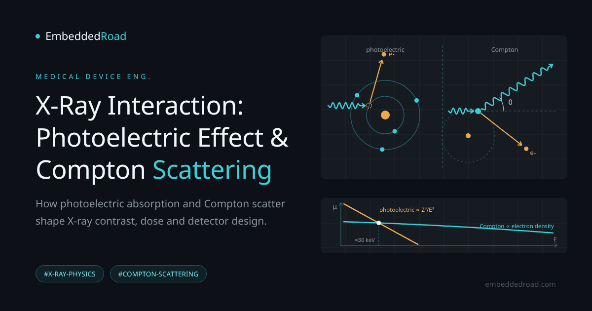
In the photoelectric effect, an incident photon is completely absorbed by a tightly bound inner-shell electron (typically K-shell), which is then ejected with kinetic energy equal to the photon energy minus the electron's binding energy. The photon ceases to exist. This is fundamentally different from scattering — there is no surviving photon to continue down the beam path, which is exactly why photoelectric absorption produces clean, high-contrast signal loss rather than redirected noise.
Threshold energy and atomic number dependence
The interaction requires the photon energy to exceed the binding energy of the shell electron, and probability spikes sharply just above each absorption edge (K-edge, L-edge) before falling off. The cross-section scales approximately as Z³ to Z⁴ divided by E³, where Z is atomic number and E is photon energy. This steep Z dependence is the single most important fact in diagnostic radiology: a small increase in effective atomic number produces a large increase in photoelectric absorption probability.
Creation of characteristic X-rays and Auger electrons
After the inner-shell electron is ejected, an outer-shell electron falls into the vacancy, releasing energy either as a characteristic X-ray photon (with energy specific to the element) or by ejecting an Auger electron. These characteristic photons have low energy and are almost always reabsorbed locally in soft tissue, so they rarely reach the detector as useful signal, but they are relevant to dose calculations and to detector materials with high-Z constituents where fluorescence escape can degrade spatial resolution.
Clinical relevance: high contrast in bone and iodine-based contrast agents
Bone (effective Z ≈ 13–14, driven by calcium) and iodine-based contrast media (Z = 53) have substantially higher atomic numbers than soft tissue (effective Z ≈ 7.4). Because photoelectric cross-section scales so steeply with Z, these materials absorb disproportionately more photons at diagnostic energies, producing the bright, high-contrast regions radiologists rely on. Iodine's K-edge sits at 33.2 keV, which is why contrast-enhanced studies are often tuned to tube voltages and filtration that place a large fraction of the spectrum just above this edge — maximizing photoelectric absorption in the contrast agent relative to surrounding tissue.
Compton Scattering: Partial Energy Transfer
Mechanism: photon collision with loosely bound outer electrons
Compton scattering is an interaction between the incident photon and a loosely bound (effectively free) outer-shell electron. Unlike the photoelectric effect, the photon is not destroyed — it transfers part of its energy and momentum to the electron and continues on a new trajectory with reduced energy. Because outer-shell electron binding energy is negligible compared to diagnostic photon energies, this interaction is nearly independent of atomic number and depends mainly on electron density.
Compton shift and scattering angle relationship
The wavelength shift of the scattered photon follows the classic Compton relation:
Δλ = (h / mₑc) × (1 − cos θ)
where h/mₑc is the Compton wavelength (2.43 pm), and θ is the scattering angle. At small angles, energy loss is minimal; at 180° (backscatter), the photon loses the maximum possible energy. In clinical energy ranges, scattered photons commonly retain 70–95% of their original energy, which means they are energetic enough to traverse tissue and reach the detector from oblique angles, carrying no spatial information about the ray path the system intended to measure.
Energy loss and recoil electron production
The energy given up by the photon becomes kinetic energy of the recoil (Compton) electron, which then deposits dose locally through ionization, similar to a photoelectric electron but without producing characteristic X-rays. This recoil electron energy is what scintillators and semiconductor detectors ultimately convert into measurable signal, regardless of whether the originating interaction was photoelectric or Compton.
Why Compton dominates at higher energies and in soft tissue
Because Compton cross-section depends on electron density rather than Z³, and because photoelectric cross-section falls off as 1/E³ while Compton falls off much more slowly, Compton scattering becomes the dominant interaction above roughly 30 keV in soft tissue and remains dominant throughout the diagnostic energy range for most tissues other than bone and contrast-filled structures. This is why soft-tissue contrast in plain radiography is comparatively flat and low — most of the attenuation differences between organs come from small density variations acting on a Compton-dominated process rather than from atomic number differences.
Comparing Photoelectric and Compton Interactions
Interaction cross-section vs. photon energy
Plotting the photoelectric and Compton components of the attenuation coefficient against energy for soft tissue shows photoelectric absorption dominating at low energies, the two crossing over at roughly 25–30 keV, and Compton scattering dominating above that. For bone and iodine, the crossover shifts upward because of the Z³–Z⁴ dependence, meaning photoelectric absorption remains clinically relevant to higher energies in these materials.
Dependence on atomic number and tissue composition
Soft tissues differ from one another mainly in electron density (mass density × Z/A ratio), which governs Compton probability, while bone, calcifications, and contrast agents differ in effective Z, which governs photoelectric probability. This distinction is precisely what CT Hounsfield units attempt to encode on a standardized scale — see CT Hounsfield Units: Tissue Density Classification for how reconstructed attenuation values are normalized against water and air to make cross-scanner comparisons possible.
Impact on image noise and scattered radiation
Compton-scattered photons that reach the detector add a diffuse background signal uncorrelated with the primary beam geometry, reducing contrast-to-noise ratio and effectively fogging the image. Photoelectric absorption, by contrast, contributes no secondary photon noise — the photon is simply gone. System designers therefore treat scatter as a noise source to be rejected, not merely attenuated.
How detector design responds to each interaction type
Detectors are engineered to maximize photoelectric absorption of the primary beam (favoring high-Z scintillators like gadolinium oxysulfide or cesium iodide) while rejecting Compton-scattered photons mainly through geometry: anti-scatter grids, air gaps, and collimation. Energy discrimination helps far less than intuition suggests, because scattered photons keep most of their energy and the primary beam is itself polyenergetic.
Clinical Impact on Image Formation
Contrast generation in radiography and CT imaging
Bone-soft tissue contrast on plain film is driven almost entirely by photoelectric differences; soft-tissue-to-soft-tissue contrast in CT relies on small Compton-dominated density differences, which is why CT requires much finer detector dynamic range and quantitative calibration than screen-film radiography ever did.
Scatter correction and beam hardening artifacts
Beam hardening occurs because photoelectric absorption preferentially removes low-energy photons from a polyenergetic beam as it traverses tissue, shifting the mean spectral energy upward and causing nonlinear attenuation that produces cupping artifacts in CT reconstructions. Scatter correction algorithms separately estimate and subtract the Compton-scattered photon contribution, often using measured or modeled scatter kernels, before reconstruction.
Optimization of tube voltage and filtration
Raising kVp shifts the balance toward Compton dominance, reducing patient dose from photoelectric absorption in bone but also reducing bone-soft tissue contrast. Added filtration (aluminum, copper) preferentially removes low-energy photons that would otherwise be absorbed photoelectrically near the skin without contributing to image formation, improving the dose-to-information ratio.
Relationship to Hounsfield unit assignment in CT
CT reconstruction converts measured attenuation coefficients into Hounsfield units referenced to water (0 HU) and air (−1000 HU). Because these values mix Compton and photoelectric contributions in energy-dependent proportions, HU values for a given tissue can shift with kVp — a practical calibration concern covered in detail in the Hounsfield units article linked above.
Detector and System Design Considerations
Scintillator and photodiode response to secondary electrons
Both photoelectric and Compton interactions ultimately produce fast electrons that excite a scintillator lattice, generating visible light photons subsequently converted to charge by a photodiode array. Scintillator thickness and density are chosen to maximize photoelectric stopping of primary photons while minimizing afterglow and cross-talk between pixels from scattered light.
Energy resolution and photon counting detectors
Photon-counting detectors using direct-conversion semiconductors (cadmium telluride, cadmium zinc telluride) can discriminate photon energy on an event-by-event basis, allowing an energy threshold set above the electronic noise floor and spectral decomposition into photoelectric-like and Compton-like components — a capability central to spectral/dual-energy CT material differentiation.
Shielding against scattered photons
Anti-scatter grids (lead or tungsten septa aligned to the focal spot) physically block obliquely incident Compton photons before they reach the detector, at the cost of absorbing some primary beam and requiring higher tube output. Air gaps and post-processing scatter estimation provide complementary, dose-neutral alternatives in some geometries.
Trade-offs between sensitivity and resolution
Thicker, denser scintillators improve photoelectric capture efficiency (sensitivity) but degrade spatial resolution through lateral light spreading, which is why fluoroscopy flat panels favor columnar cesium iodide, whose needle-like crystals channel light toward the photodiodes. Every detector design decision — material, thickness, grid ratio, energy threshold — is a negotiated trade-off between maximizing useful photoelectric-driven signal and minimizing Compton-driven scatter noise, and the correct balance depends on the specific clinical task the system is built for.
Learning
Sign in to track your progress.
Evidence
Public projects engineers linked to X-Ray Interaction: Photoelectric Effect, Compton Scattering.
No engineer has linked a project to this topic yet. Built something that proves it? Add the project and tag it with medical-devices-x-ray-interaction-photoelectric-effect-compton-sca — it then shows here and on your public profile.